emfret ANALYTICS公司主营业务:抗体(心血管研究类),网站:http://www.emfret.com/
Flow cytometric analysis of mouse platelet surface glycoproteins
- Preparation of diluted or washed whole blood
- Preparation of washed mouse platelets
- Single-color analysis of platelet surface glycoprotein expression
- Two-color analysis of platelet activation
Immunohistochemistry (with acetone-fixed frozen sections)
Immunoblot (Western Blot Analysis)

Buffers and reagents
Tris buffered saline/Heparin (TBS/Hep): 20 mM Tris/HCl; 137 mM NaCl; pH 7.3; containing 20 U/ml Heparin.
Phosphate buffered saline (PBS): 137 mM NaCl; 2.7 mM KCl; 1.5 mM KH2PO4; 8 mM Na2HPO4; pH 7.14.
Tyrode’s Buffer (modified): 134 mM NaCl; 0.34 mM Na2HPO4; 2.9 mM KCl; 12 mM NaHCO3; 20 mM Hepes; pH 7.0; 5 mM glucose; 0.35% (w/v) bovine serum albumin
Apyrase: 10 U/ml stock in H2Obidest (stored at -20°C)
Prostacyclin (Prostaglandin 2, PGI2): 1 mM stock in H2Obidest (stored at -20°C)
CaCl2: 1 M stock in H2Obidest
Preparation of diluted or washed whole blood
Take 50 µl blood with a heparinized glass capillary (e.g. ringcap) from the retro-orbital plexus into 1.5 ml tubes containing 200 µl of TBS/Heparin (20 U/ml).
Dilute in 1 ml of Tyrode´s buffer and use for flow cytometric analysis.
To prepare washed blood, centrifuge the diluted blood at 900 x g for 5 min, remove the supernatant and resuspend the pellet in 1.25 ml Tyrode´s buffer.
Add 1 mM CaCl2 directly before the start of the experiment.
Note: For thrombin-induced platelet activation, plasma has to be removed to prevent anti-thrombin activity
Preparation of washed mouse platelets
The preparation of washed platelets is a more time consuming method, that requires a larger amount of blood, but yields a platelet preparation that can be used for several hours.
Collect 0.5 - 1 ml blood with 1.5 cm glass capillaries from the retro-orbital plexus into a 1.5 ml tube containing 200 µl of TBS/Heparin (20 U/ml).
Centrifuge the sample for 5 min at 500 x g and transfer the platelet rich plasma (PRP) into a new tube. For best recovery of platelets, take the complete white phase including some red blood cells.
Centrifuge the platelet suspension for 8 min at 300 x g, and transfer the PRP without any red blood cells into a new tube.
Add 0.5 µM prostacyclin (PGI2) and centrifuge at 1300 x g for 5 min.
Resuspend the platelet pellet in 1 ml Tyrode’s buffer, add 0.02 U/ml apyrase and 0.5 µM prostacyclin, incubate for 5 min at 37°C, and centrifuge for 5 min at 1300 x g.
Repeat step 5 and resuspend the platelet pellet in 0.5 ml Tyrode’s buffer, add 0.02 U/ml of apyrase and incubate for 30 min at 37°C.
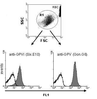
Single color analysis of platelet surface glycoproteins
Combine 5 µl of specific or negative control antibodies and 25 µl diluted whole or washed blood in the assay tube and vortex mix for 1-2 seconds.
Incubate for 15 min at room temperature.
Stop reaction by addition of 400 µl PBS and analyze within 30 min.
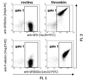
Two color analysis of platelet activation
Add 5 µl specific or negative control antibodies to the assay tube together with 5 µl of a pan-platelet marker.
At this point, any additional reagents (e.g. inhibitors) are added in small volumes (< 5 µl).
Add 26 µl diluted whole or washed blood or washed platelets (1 million) and vortex mix for 1-2 seconds.
Incubate for 15 min at room temperature. Stop reaction by addition of 400 µl PBS and analyze within 30 min.
Please note: It is not recommended to perform immune staining on (para-)formaldehyde-fixed samples, since most rat antibodies yield only poor results under these conditions on mouse tissues.
Mount sections from frozen tissue on slides and fix in ice cold 100% acetone for 20 min at 4°C. Dry samples over night at RT.
To minimize reagent volumes, a hydrophobic ring may be drawn with a water repellent pen around the tissue section on the slide. During all following procedures, this encircled area should not dry up. Therefore, it is recommended to perform incubation steps in a humid box.
Incubate slides in PBS for 20 min.
When using peroxidase-conjugated secondary antibodies, incubate slides with 0.03% H2O2 for 10 min at RT to inhibit endogenous peroxidase activity. Rinse slides three times in PBS for 5 min each wash.
Incubate slides for 1 hour at RT in blocking solution (serum from host species of secondary antibody to be used), diluted 1:10 or 1:100 in PBS.
Cover slides with primary antibody (2-10 µg/ml) in blocking solution and incubate for 2 h at RT.
Blot excess liquid from slides and rinse three times in PBS for 5 min each wash.
Cover slides with fluorophore- or peroxidase-conjugated secondary antibody (e.g. affinity purified donkey anti-rat IgG-FITC or -HRP, Jackson ImmunoResearch, West Grove, PA) diluted in blocking solution according to the manufacturer’s instructions. Incubate for 1 h at RT.
Blot excess liquid from slides and rinse three times in PBS for 5 min each wash.
a) Fluorescently stained samples can be directly analyzed by fluorescence microscopy.
b) For visualization of peroxidase-labeled structures, cover slides with freshly prepared 3-amino-9-ethylcarbazole (AEC) substrate and incubate until red staining is visible under the microscope, time can vary between 5 and 30 min. Stop reaction by rinsing with deionized water before orange background staining occurs. For counterstaining, incubate samples for 30-240 s with hematoxylin and rinse slides for 20 min with running warm water. Mount sections with aqueous embedding solution (e.g. Aquatex, Merck).
Buffers and Reagents
PBS/EDTA: 137 mM NaCl, 2.7 mM KCl, 1.5 mM KH2PO4, 8.0 mM Na2HPO4 x 2 H2O, pH 7.3; 5 mM EDTA
IP buffer: 40 mM Tris/HCl, 0.3 M NaCl, 1 mM EDTA, 2 mM PMSF, 10 µg/ml Leupeptin, 10 µg/ml Aprotinin, 1 µg/ml Pepstatin
Igepal: 10% stock in H2Obidest
2x SDS-sample buffer: 10% beta-mercaptoethanol (for reducing buffer), 10% Tris buffer (1.25 M), 20% Glycerin, 4% SDS, 0.02% Bromophenolblue
Wash mouse platelets or cells (10 million per immunoprecipitation) 3 x in1 ml PBS/EDTA (5 min, 1300 x g) and resuspend in IP-buffer.
Lyse platelets by addition of 1% Igepal for 15 – 20 min at RT.
To remove cell debris, spin 5 min at 10,000 x g and transfer supernatant to a new cup.
Preclear by adding 20 µl of washed protein G agarose (PGA). Incubate with rotation for at least 3 h at 4°C.
Centrifuge the samples for 5 min at 3000 x g. Fill supernatants into new tubes, add the antibody at a concentration given in the data sheet (usually between 2 and 5 µg/ml), and after 30 min add 25 µl of washed PGA. Incubate with rotation for at least 2 h at 4°C, preferably over night.
To wash the precipitate, centrifuge the samples for 1 min at 3000 x g. Add 1 ml of 1 x IP buffer containing 1% NP-40 to the PGA pellet. Centrifuge for 1 min at 3000 x g.
Wash the PGA pellet twice in plain IP-buffer (1 min at 3000 x g).
Resuspend the PGA pellet in 25 µl of 1x SDS-sample buffer (reducing or non-reducing) and boil for 5 min. The samples can then be frozen (-20°C) for later use, or SDS-PAGE and subsequent immunoblot analysis can be performed.
Buffers and reagents
Lysis buffer: 40 mM Tris/HCl, 0.3 M NaCl, 1 mM EDTA, 0.05% NaN3, 1% NP-40
PBS: 137 mM NaCl, 2.7 mM KCl, 1.5 mM KH2PO4, 8.0 mM Na2HPO4 x 2H2O, pH 7.3
PBS-5% milk: PBS containing 5% non-fat dry milk
PBS/T: PBS containing 0.1% Tween 20
PBS/T-1% milk: PBS/T containing 1% non-fat dry milk
All steps can be performed at room temperature:
1. Prepare lysate of mouse platelets or cells in lysis buffer and incubate with (2x) SDS sample buffer for 5 min at 95°C.
2. Separate proteins by SDS-polyacrylamide gel electrophoresis (SDS-PAGE) under reducing or nonreducing conditions, as indicated in the respective data sheet, and transfer the proteins to PVDF membrane.
3. Block the membrane with PBS-5% milk for 1 h with agitation.
4. Incubate the membrane with 2-5 µg/ml of primary antibody in PBS/T-1% milk for 1 h with agitation.
5. Rinse the membrane twice with PBS/T followed by two 30 min washing periods in PBS/T. Washing procedure may be reduced to three periods of 10 min each, but that should be determined individually.
6. Incubate with secondary antibody (HRP-conjugated anti-rat Ig, e.g. from DAKO, Glostrup, Denmark, #P0162) in PBS/T-1% milk
for 45–60 min with agitation.
7. Rinse the membrane twice with PBS/T followed by two 30 min washing periods in PBS/T.
8. Visualize bound antibody with ECL (enhanced chemiluminiscence).
技术支持:易动力网络